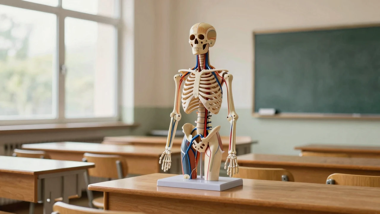
To tell you the truth, when I first moved this 85-centimeter neurovascular skeletal model into anatomy class, I actually had a pretty bad heart. After all, they used to rely on PPT and wall charts, and students looked at blood vessels walking like mazes, remembering and forgetting. As a result, the whole classroom atmosphere changed.

The most intuitive feeling is...——Save your mouth.I don't know. I used to say, "The artery's on the right side of the nerves." I had to do it three times on the blackboard. Now let the students come around, point at the red artery and yellow nerves on the model, and say, "Look, they're going right here." Half a minute, the students' eyes were bright.
The best design for this model is "half-side color." The left is pure bones, the right is an vascular nerve. I let two groups of students observe each: one on the right side, and the other on the left side only. Exchange of information in five minutes and co-certification. It worked so well.——Because students have to think about where this nerve comes from, not passively.
I put the model in the middle of the classroom, and each group gave a colored pen and a transparent film. The task is simple: draw the arteries, veins and nerves on the right side of the model on the translucent membranes, and then cover the skeletal matching on the left side. A girl drew and cried out: "The original neurological is from the back of the gill!——This "self-discovery" thing works 10 times more than a teacher.
I occasionally deliberately turn the model in a direction where students sneak a colored hose behind closed eyes (the vascular nerve on the model is softly moved) and then open their eyes to change. This is particularly effective for refresher studies, where students, in order to win, wish to carve the starting point of every vein into their minds.
I made a mistake at first.——Call all the students together before the model. Speak.I don't know. As a result, the rear row was completely invisible, and the front was sidelined. Later, it was changed to "Small Liners": each time only 5-6 people came round, the next set was changed in five minutes. Other students use tablets for 3D anatomical pre-learning, just in time.
And...Don't be greedy.I don't know. One session focused on two or three critical areas, such as "Neurovascular beams of upper limbs" or "The artery of lower limbs". Models are repeated and students cannot remember the information.
Seriously, ever since I used this model, I've found myself having less time to prepare. Because it's too intuitive, and there are many details that need to be explained over and over again, the students can see it at a glance. Now, every time I go to class, I set up the model ahead of time, and I sneak up on the "whoa."——That sense of accomplishment is better than a paper.
Participation in discussions
The model does smell good, and used to say that the nervous walker's skin had torn a student's face.
It's kind of a fun game, but isn't it easier to move more soft pipes?🤔
It's hard to see in the back row. Rotation is the right thing to do.
Wow, the students found their own nerve around there, and they're so excited.