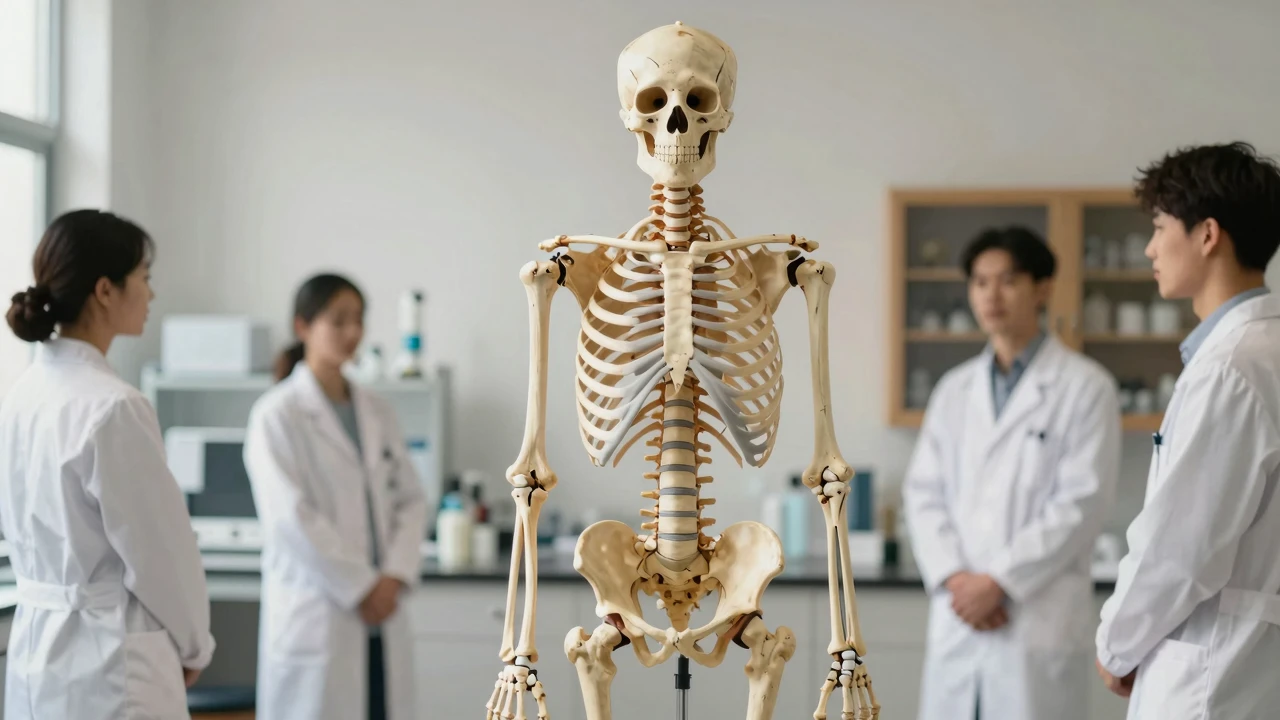
In the anatomy lab in the school building, a model of 175 cm, marked by the neurological root, is being surrounded by a resident. It is not a mere decoration, but a three-dimensional structure of anatomy is moved directly to the clinic to help the doctor "preprepare" the direction of each vertebrae before the operation.

In the rotation of an osteoporator, "surgery simulations" can be performed first on models for common intervertebral discs, fractures or congenital malformations. Radiologists use models to compare the cut side of X-rays, CT or MRI to quickly locate the location of the abnormal vertebrae. In the face of multiple trauma, young doctors in emergency care often reset the model to confirm whether a plethora or pelvis injury was involved.
A multi-centre comparative study published in 2023 by the Chinese Osteroid Journal showed that the interpretation accuracy of vertebrae fractures increased from 78 per cent to 92 per cent, with an average time reduction of about 15 minutes. The spinal surgery team at a Beijing hospital reported a decrease of approximately 12 per cent in surgical haemorrhage and 1.3 days in hospital days following pre-operative planning using the model.
Despite the obvious advantages of physical models in space perception, production costs remain a key factor limiting access. As the cost of 3D printed materials declines year by year, it is expected that in the future an exclusive model tailored to patient-specific anatomy data will be achieved. At the same time, the translation of model training results into algorithms for video diagnostics is the direction of interdisciplinary cooperation.
Would it make clinical decision-making more like an accurate puzzle game by moving anatomy 'hardware' into the clinic?
Participation in discussions
This nerve root is more clearly marked than a dead-end back.
175cm is standard height? What if you meet a very tall or short patient?
That's the kind of face that can be opened up.
Just looking at the data, I'd like to know how much this model is worth.
If you can print the patient's specific CT data directly for pre-operative simulation, you'll have to do with your brain now.